 Characterizing Cell and Tissue Mechanical Properties
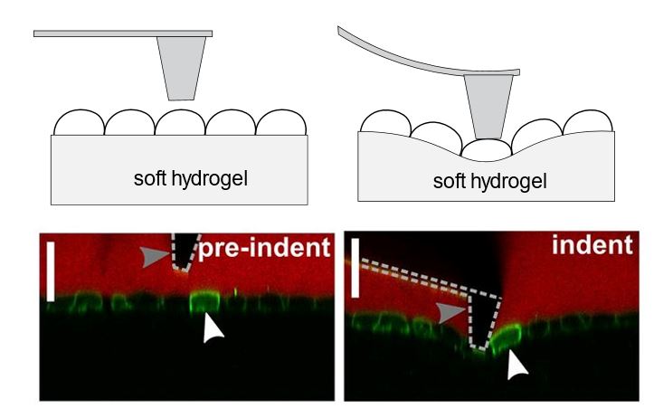
Sustaining and generating mechanical forces is a normal part of physiology for mammalian tissue: Intestinal epithelia are stretched during peristaltic movements in the gut, lung alveoli deform during breathing and skin epidermal layers provide a mechanical barrier to the external environment. Mechanical integrity and force transmission in even the simplest tissues is critical to their proper function. This is particularly apparent in diseased states where point mutations to cytoskeletal or junctional proteins result in tissue mechanical failure such as blistering, cracking and hemorrhaging. Characterizing the mechanical properties of single cells and simple tissues from a materials science and engineering perspective could lead to a better understanding of diseases of cell and tissue fragility. The lab is currently interested in understanding the mechanical properties of cells in a number of different systems and tissues including the kidney, gut, skin and blood.

Harris, A.R., Daeden, A. and Charras, G.T., 2014. Formation of adherens junctions leads to the emergence of a tissue-level tension in epithelial monolayers. J Cell Sci, 127(11), pp.2507-2517.
 Engineering Protein, Cell and Tissue Mechanical Properties for Regenerative Medicine
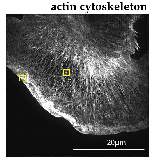
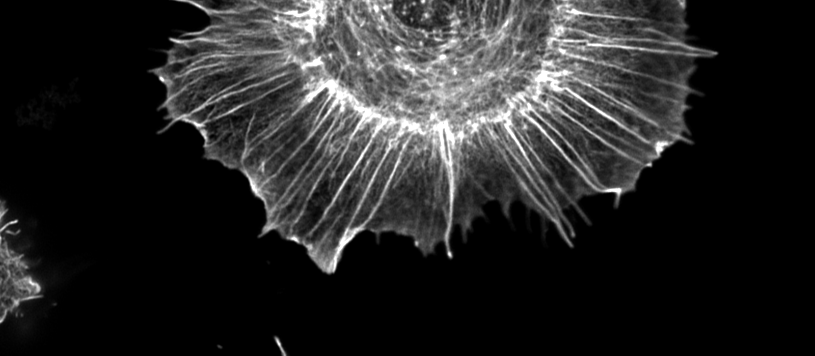
One of the major determinants of cell mechanical properties is a filament forming protein called actin. Actin filaments are assembled into distinct higher order structures (including bundles, meshes and cables) by an array of specialized binding and crosslinking proteins. The organization of actin filaments within each structure is directly linked to its function through the ability to generate specific mechanical forces. For example, aligned bundles of actin filaments in stress fibers pull on the extracellular matrix and dendritic actin networks push the leading edge of migrating cells forwards. Missense mutations to actin crosslinking proteins are associated with a number of diseases including familial Focal Segmental Glomerulosclerosis (FSG), Periventricular nodular heterotopia (PVNH), Myofibrillar and Distal Myopathies, many of which present with symptoms associated with compromised cellular force generation and tissue fragility. Understanding how actin filaments are organized into distinct force generating structures through the activity of filament crosslinking proteins is therefore interesting and important from both a biophysical and clinical perspective. Despite this, classification of subcellular actin structures is currently based on user identification from diffraction limited microscopy data, which is insensitive to subtle changes in organization and convoluted with cell to cell variations in geometry. This limits not only our understanding of the mechanisms of disease but our ability to develop new therapeutic strategies. The lab takes a multidisciplinary approach to understanding how actin filaments are organized into force generating structures using tools such as machine learning for image analysis and purification of actin regulatory proteins to characterize their behavior in vitro.

Harris, A.R., Jreij, P., Belardi, B., Joffe, A.M., Bausch, A. & Fletcher, D. A. 2020. Biased localization of actin binding proteins by actin filament conformation. Nature Communications 11, 5973 (2020).
Harris, A. R. Belardi, B., Jreij, P., Wei, K., Bausch, A. & Fletcher, D. A. 2019. Steric regulation of tandem calponin homology domain actin-binding affinity. Mol. Biol. Cell mbc. E19-06-0317.
 Engineering Tools to Study Biology
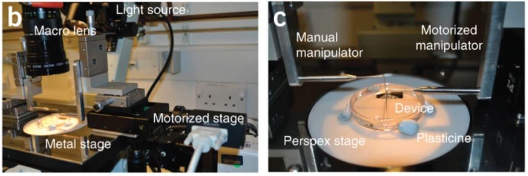
The length scale of a single cell or simple tissue is on the order of tens to hundreds of micrometers. In order to study the mechanical properties of samples on this length scale we can adapt and shrink down common tools for mechanical testing. Additional constraints on making these measurements are that the sample is alive and needs to be kept in conditions that are as close to physiologically relevant as possible. Engineering tools for studying biological systems presents an intriguing problem and often requires creative solutions. The lab uses established techniques such as Optical Microscopy and Atomic Force Microscopy but also develops new tools that provide quantitative measurements of the behavior of biological samples in a controlled environment, across a range of length scales.

Harris, A.R., Bellis, J., Khalilgharibi, N., Wyatt, T., Baum, B., Kabla, A.J. and Charras, G.T., 2013. Generating suspended cell monolayers for mechanobiological studies. Nature protocols, 8(12), pp.2516-2530.
 Engineering Education
Creating an inclusive environment in the lab and the classroom is paramount for success. The CTE lab has active research projects in the area of engineering education and developing novel devices for laboratory experiments and experiential learning.
 Collaborators
Impact Research Lab
MuBEST Lab
https://carleton.ca/mubest-lab/
Maganti Lab
Willmore Lab
https://carleton.ca/willmorelab/
Materials as Machines Lab
https://carleton.ca/materialsasmachines/
Tissue Engineering and Bioimaging Lab
https://carleton.ca/mostacoguidolin/research/hub/
Tissue Engineering and Applied Materials Hub

https://www.teamhubottawa.com/
 Funding


