Field Techniques
Our test involved transferring all high-resolution images in the camera memory card to a computer. For field trials, we used the same general method, and counted pigment particles later in the lab because of limited field time and the need to reconfirm. In future, we plan to use a live-feed camera and direct USB hookup to collect particles for AMS analysis. This is to confirm that they really are pigment, which is highly likely. We can do this by rephotographing a lab spatula placed alongside, until we pick up the particle. Another photo should confirm its extraction.
In the field, we screened each 5 mm peel, collecting, labelling, and bagging any organic material in it for dating. Only one of ten sites had sterile sand. Typically, we got 50 to 100 peels in 50 cm of soil, many with AMS samples. AMS actually counts carbon atoms in a milligram sample and is more realistic than conventional carbon-dating, which demands a gram sample.
Enhancing Pictographs with High Resolution Photography
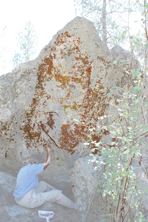
Motif photos are unneeded for dating because their colour has faded. Our pigment-coated paper on a newly-scraped surface is preferred. However, we recorded the art to later enhance hidden motifs even on a blank wall. Pigments to a depth of 8mm in sandstone appear in photos like Las Vegas lights on a Saturday night.
Actual Field Tests
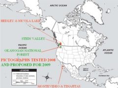
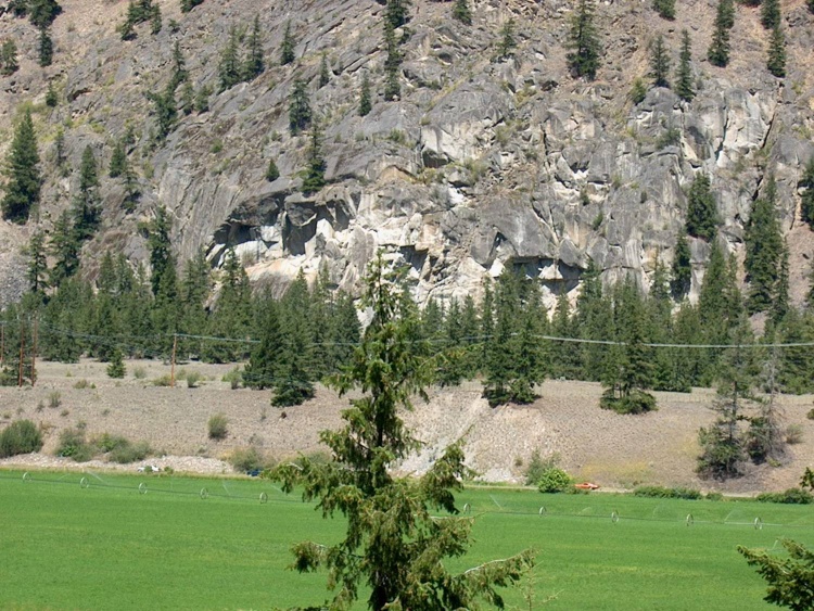
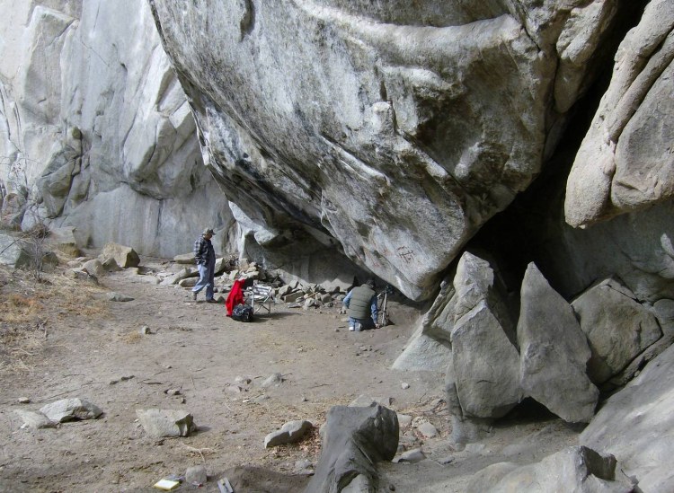
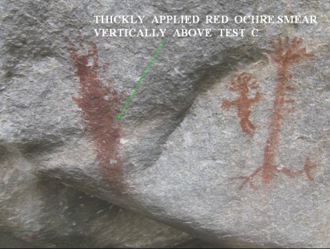
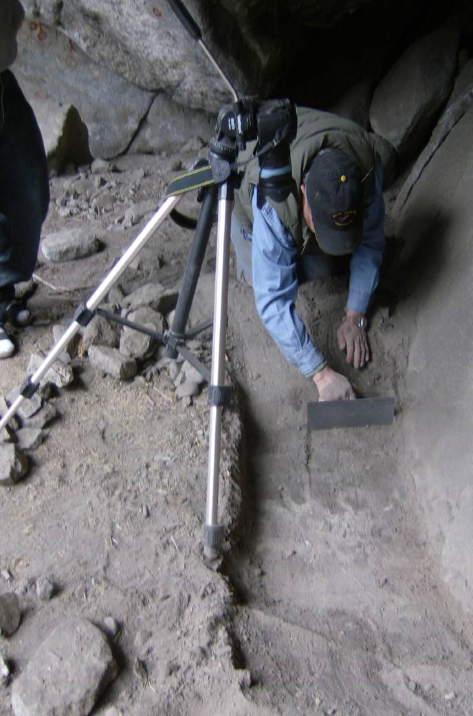
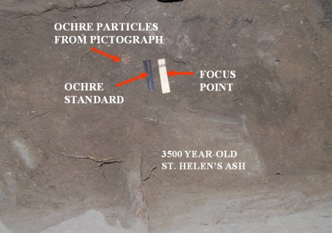
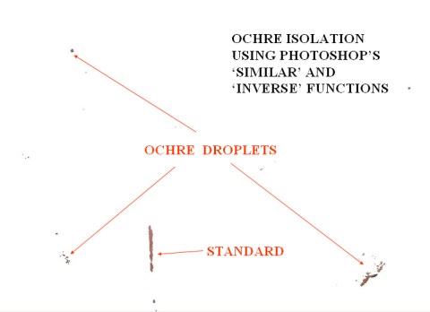
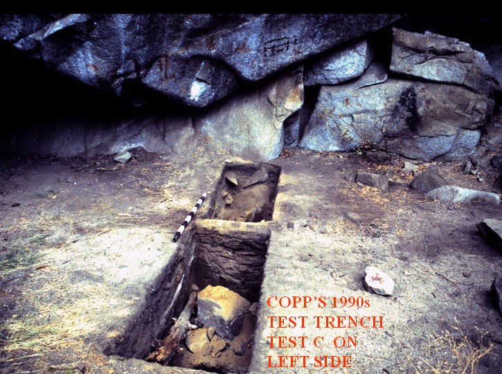
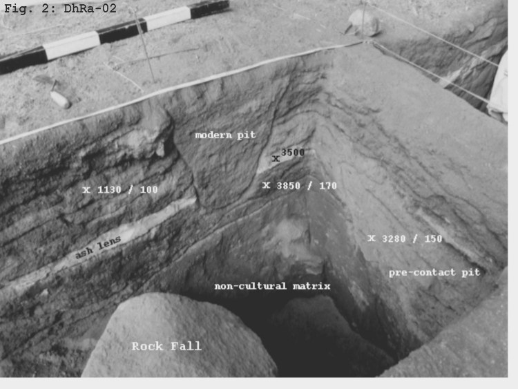
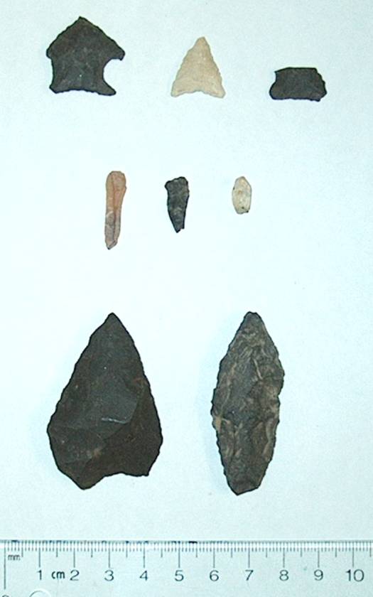
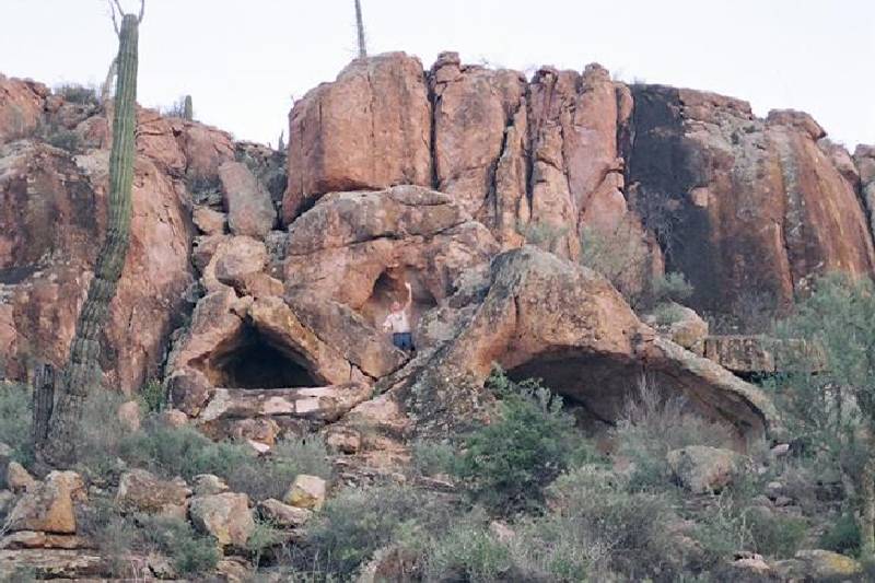
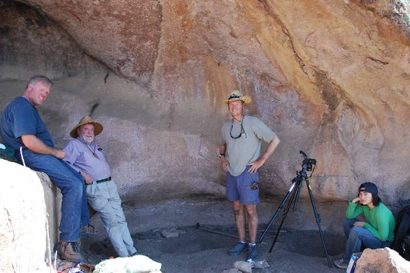
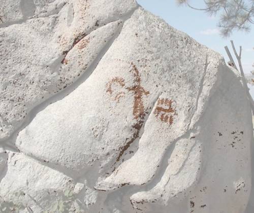
Our first field trial was at the Hedley rockshelter in British Columbia where I unsuccessfully tried automatic camera focussing. Sand grains were unclear, so I switched to manual focus. (Slide 11) The rockshelter was also in shadow, so we standardized our light source to the camera flash (Slide 12). Three mid-shelter tests in the dry ashy soil were chosen on the basis of soil depth below the art measured with a spike or knitting needle. The best test was at C between A and B, and where the tripod stands. (Slide 13) A pebble dropped from a repeatedly-painted band or palimpsest, rather than a single-painted motif, enhanced our chances of finding where droplets fell. (Slide 14) We outlined a rectangle on the soil surface and scraped 5 mm peels or levels within it down to sterile soil or bedrock. C produced peels with plant, beetle and bone fragments for dating and identification to 450 mm subsurface. (Slide 15) Visible red ochre at the bottom on volcanic ash overlay fallen rock. Other particles are rendered visible under software enlargement and its Inverse function (Slide 16). This peel was cross-dated with the same level 1 m away in Copp’s now-infilled test-trench (Slide 17). In the 1990s, he excavated 5 contiguous m2 units to a depth of 1m, reaching the well-dated 3500 year-old St. Helen’s volcanic ash. (Slide 18) Ochre fragments in his upper 20 cm were AMS-dated on bone to 1130 years ago, while lower bone samples dated 3280 and 3580 years ago, the latter below the ash. (Slide 19) His sparse lithics and abundant bone fragments, fire-cracked rock and rolled birchbark, emphasized the ceremonial rather than practical nature of the site.
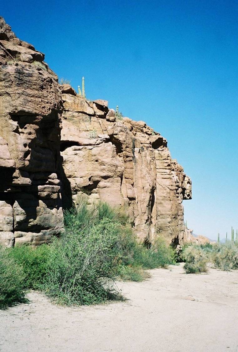
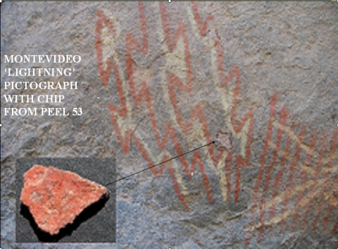
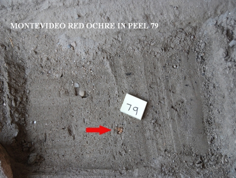
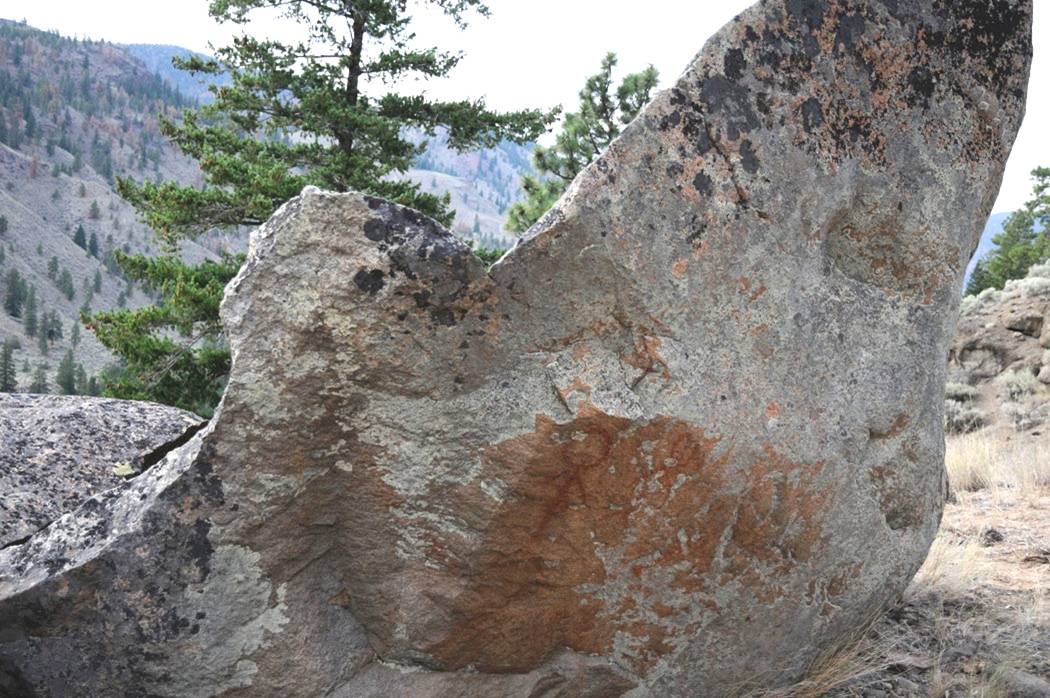
In April 2008, I led an expedition to Baja California to the sites of Montevideo and Las Tinajitas. (Slide 21) Variable sunlit walls required a canopy for shade and a flash to keep our reflected RGB values constant. At Montevideo (Slide 22) we tested under the ‘lightning bolt’ pictograph below the main painted wall. We scraped 131 peels to a depth of 600 mm, encountering an ochre-coloured rock chip from the pictograph at 180 mm. Charcoal, plant, shell, bone or stone fragments were found in most levels. Enlarging revealed ochre particles at 125mm above the painted chip. As the chip must have fallen after the pictograph was painted, any AMS date below the chip would better date the motif (Slide 23). A week later, we tested two shelters at Las Tinajitas about 75 km away (Slide 24). We tested twice in the Lower Shelter (Slide 25). Note the smeared ceiling art above the crew.
In July, we tested boulder pictographs in the Nicola Valley of British Columbia, 5 of which had underlying soil. (Slide 26) At Canford Bird Lady, we scraped 23 peels under thick leaf litter, 7 of which had ochre particles and AMS samples. Lower peels 10, 14 and 17 had yellow, red and yellow & red ochre particles at depths of 110, 130 and 145 mm, respectively, along with AMS samples. (Slide 27) The west side of Monck Rock produced 28 peels to a depth of 405 mm, with only peel 5 at 60 mm having red ochre particles and datable leaves. Monck East on the other side of the boulder with thick surface leaf litter, had red ochre in peels 2, 13 and 14 at 72, 312 and 330 mm, the latter two recommended for dating. At 10 Mile Woman, we scraped 20 peels to a depth of 200 mm. (Slide 28) This is a menstrual site depicting spruce boughs used to cover the girl in her coming-of-age rites. Peel surfaces 8, 12, 15 and 16 at depths of 75, 95, 110 and 115mm had red ochre particles, with only peel 8 having datable twigs, it and the other three peels having questionable root contamination.
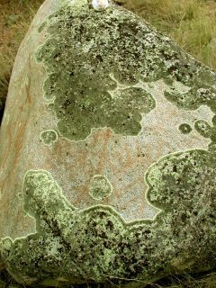
(Slide 29) Lichens are gradually creeping over the art at Nicola. There, as in other places subject to public viewing, officials are under pressure to expose the art by removing lichen. Any such lichen removal is not advised because it damages the art. Subjects of future papers will be penetrating the lichen photographically and enhancing the underlying art, plus AMS dating of pictographs in the Okanogan Valley of Washington State.
Slide 29
Conclusions
My method demands sand or soil under wall art and sufficient natural or cultural organic material in the soil for AMS dating, But it can overcome the problems associated with existing methods without destroying the pictograph or soil layers beneath it.